|
The 3D heart at the right can be rotated using the mouse to view the arrhythmia in more detail. |
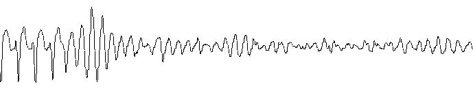
ECG rhythm strip, Lead II, 50 mm/sec, 10mm/mV. The beginning of this ECG shows a run of rapid ventricular tachycardia (VT) that degenerates into ventricular fibrillation (VF), characterized by a low-amplitude, chaotic baseline where no distinct waveform pattern can be identified.