The underlying cause of many arrhythmias is
the development of a reentrant circuit of electrical activity
that repetitively stimulates the heart and produces contractions at a rapid rate.
During tachycardia, a single wave can rotate as a spiral wave,
producing fast rates and complex wavefoms. During fibrillation, a single spiral wave
can degenerate into multiple waves that are no longer spatially coordinated. Because the spatial pattern of contraction is determined by
the spatial pattern of electrical waves, hearts in arrhythmia can no longer
pump blood effectively and in some cases may cause potentially lethal cardiac arrest.
To learn more about sinus rhythm and several of the most common types of arrhythmias,
click on the tabs above. Each rhythm is demonstrated using a movie, sample ECG, and
three-dimensional interactive simulation.
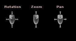
The contracting three-dimensional heart on the left can be rotated, zoomed, and panned by
clicking and moving the mouse as indicated.
During normal rhythm (also called sinus rhythm), the heart beats regularly, producing
a single coordinated electrical wave that can be seen as a normal
electrocardiogram (ECG).
During arrhythmias such as ventricular tachycardia and
ventricular fibrillation, this normal behavior is disrupted, and the ECG
records rapid rates with increased complexity.