Movies

This section contains experimental movies of single cells:
Contracting cells   and   Calcium waves in cells    

Most cardiac cells are approximately cylindrical, with a length of around 100 microns and diameter of 10-15 microns.
Each cell connects to roughly 8-9 neighboring cells, with the sites of these connections preferentially located at the ends of the long axis.
Cells connected at their ends form long cardiac fibers that wrap around the heart and together generate sufficient contractile force to pump blood.
Calcium concentrations inside the cell respond to electrical stimulation and initiate the mechanics of contraction.

Cardiac cells (size ~ 10x100 microns)

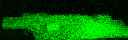
Contracting ventricular cell
Format: gif movie(367K)


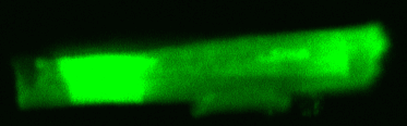
Contracting ventricular cell
Format: gif movie(397K)


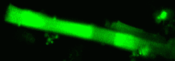
Contracting ventricular cell
Format: gif movie(674K)


Calcium imaging in cardiac cells


Intracellular calcium waves shown using fluo-4 dye (which fluoresces in proportion to the calcium concentration) and confocal microscopy
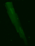
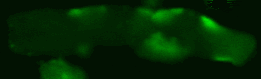
Propagating calcium wave eliciting
contraction of a ventricular cell
Format: gif movie(357K)


Propagating calcium wave eliciting
contraction of a ventricular cell
Format: gif movie(138K)


Propagating calcium waves
from multiple sites
Format: gif movie(133K)


Propagating calcium wave
from one site
Format: gif movie(430K)


Propagating calcium waves
Format: gif movie(324K)


Multiple calcium sparks
Format: gif movie(292K)